Dr. Alptekin Özkoç

STRUCTURAL HEART INTERVENTIONS

WHAT IS TAVI?

TAVI

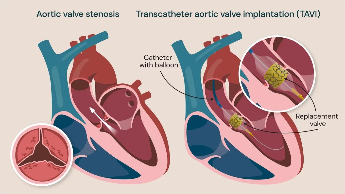
TAVI, or Transcatheter Aortic Valve Implantation, is a minimally invasive treatment for severe aortic stenosis.

Instead of replacing the valve through open-heart surgery, a new valve is delivered using a catheter, most commonly through the femoral artery.

The new valve is positioned inside the diseased native valve and expanded into place.

TAVI can be performed without opening the chest and without the need for cardiopulmonary bypass.

Depending on the patient's condition and the center's protocol, the procedure may be performed using conscious sedation or general anesthesia.

The decision between TAVI and surgical aortic valve replacement should be individualized based on anatomy, age, surgical risk, life expectancy, comorbidities, and patient preference.

The procedure typically takes 60-90 minutes, and patients are generally discharged from the hospital 2-3 days later
WHAT IS MITRACLİP?

MitraClip

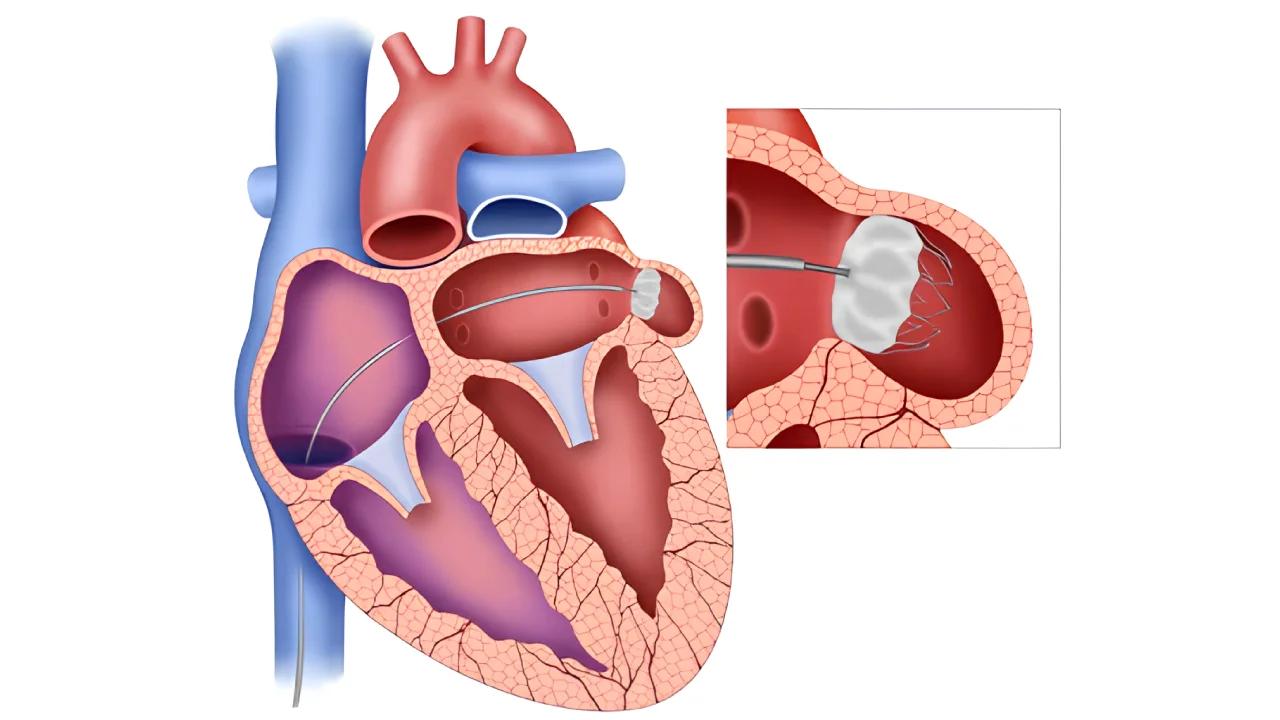
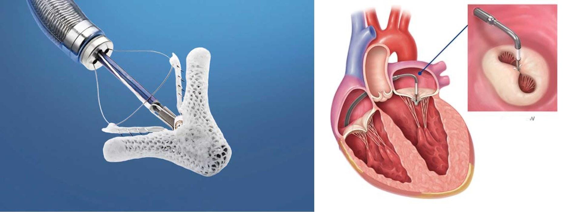
MitraClip is a minimally invasive, catheter-based treatment for significant mitral regurgitation (MR), a condition in which the mitral valve does not close properly and allows blood to leak backward into the left atrium.

During the procedure, a small clip is delivered to the mitral valve through a catheter inserted through a vein in the groin. The clip grasps the mitral valve leaflets and brings them closer together, helping to reduce the amount of blood leaking through the valve.

Who May Benefit?

MitraClip may be considered for selected patients with significant mitral regurgitation, particularly those who:

  • Have significant symptoms
  • Have suitable mitral valve anatomy
  • Are at increased risk for conventional surgery
  • Have functional or degenerative mitral regurgitation when TEER is appropriate

Potential Benefits

  • Reduces mitral regurgitation
  • May improve shortness of breath
  • May improve exercise capacity
  • May improve quality of life
  • Avoids open-heart surgery in appropriately selected patients

Key Message

MitraClip, also known as Transcatheter Edge-to-Edge Repair (TEER), offers a minimally invasive treatment option for selected patients with significant mitral regurgitation. The suitability of the procedure depends on the type and severity of mitral valve disease, valve anatomy, heart function, and the patient's overall clinical condition.

The procedure typically takes 1-2 hours and patients are generally discharged from the hospital the following day.

WHAT İS TRİCLİP?

TriClip

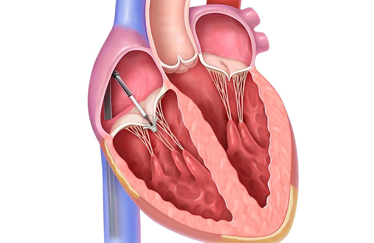
TriClip is a minimally invasive, catheter-based treatment for tricuspid regurgitation (TR), a condition in which the tricuspid valve does not close properly and allows blood to flow backward into the right atrium.

During the procedure, a small clip is delivered to the tricuspid valve through a catheter, usually inserted through a vein in the groin. The clip grasps the valve leaflets and brings them closer together, helping to reduce the amount of blood leaking through the valve.

Who May Benefit?

TriClip may be considered for selected patients with significant tricuspid regurgitation, particularly those who:

  • Have significant symptoms
  • Have suitable tricuspid valve anatomy
  • Are at high risk for conventional surgery
  • Remain symptomatic despite appropriate medical treatment

Potential Benefits

  • Reduces tricuspid regurgitation
  • May improve shortness of breath and fatigue
  • May reduce leg swelling
  • May improve exercise capacity
  • May improve quality of life
  • Avoids open-heart surgery in appropriately selected patients

Key Message

TriClip, also known as Transcatheter Edge-to-Edge Repair (T-TEER), provides a minimally invasive treatment option for selected patients with significant tricuspid regurgitation.

The procedure typically takes 1-2 hours, and patients are generally discharged from the hospital the following day.

WHAT İS TRİCVALVE?

TricValve

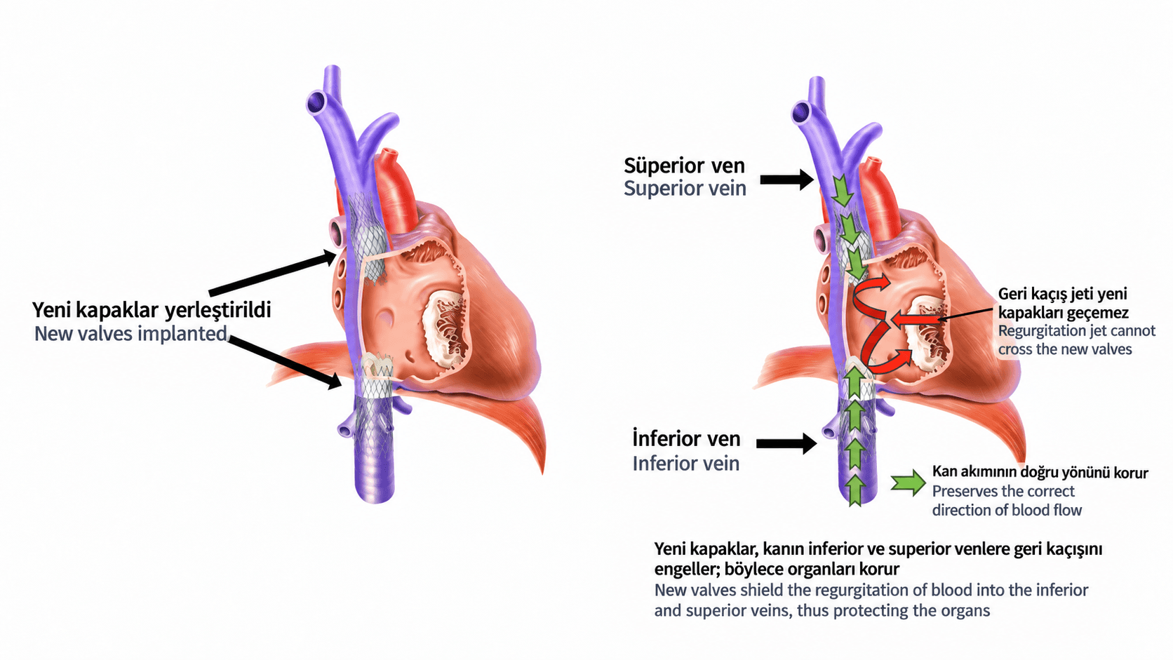
TriValve is a minimally invasive, catheter-based approach designed to treat selected patients with severe tricuspid regurgitation (TR) who are at high risk for conventional surgery.

Instead of directly repairing or replacing the tricuspid valve, the procedure involves placing transcatheter valves in the inferior vena cava (IVC) and/or superior vena cava (SVC).

These valves help prevent the backward flow of blood caused by severe tricuspid regurgitation, reducing venous congestion and pressure in the systemic veins.

Who May Benefit?

TriValve may be considered in selected patients with:

  • Severe tricuspid regurgitation
  • Significant right-sided heart failure symptoms
  • Severe systemic venous congestion
  • High or prohibitive surgical risk
  • Anatomy suitable for caval valve implantation

Potential Benefits

  • Reduces backward venous blood flow
  • Decreases systemic venous congestion
  • May reduce leg and abdominal swelling
  • May improve symptoms and quality of life
  • Provides a minimally invasive option for patients who are not suitable for conventional surgery

Key Message

Caval valve implantation (CAVI) is an emerging transcatheter approach for selected patients with severe tricuspid regurgitation and advanced venous congestion.

The treatment strategy depends on the patient's anatomy, severity of tricuspid regurgitation, right ventricular function, pulmonary pressures, and overall clinical condition.

The procedure typically takes 60 minutes, and patients are generally discharged from the hospital the following day.

What Is Left Atrial Appendage Occlusion (LAAO)?

Left Atrial Appendage Occlusion (LAAO)

Left atrial appendage occlusion (LAAO) is a non-surgical procedure that closes a small pouch in the heart's left atrium, particularly in patients with atrial fibrillation (AF). During AF, blood can pool in this area and form clots, which can travel to the brain and cause a stroke.

Why Is It Done?

Its main purpose is to reduce the risk of stroke.

It is an alternative treatment option for suitable patients who need long-term blood thinners but have a high bleeding risk, cannot take blood thinners, or have a history of serious bleeding.

How Is It Done?

The procedure is usually performed by entering through a thin catheter from an artery in the groin. The catheter is advanced to the left atrium through a special passage from the right side of the heart, and a special closure device is placed inside the left atrial appendage.

During the procedure, intracardiac imaging methods are used to confirm the device is in the correct position.

  • The procedure takes about 1 hour.
  • It is usually performed under anesthesia or sedation.
  • Most patients are monitored in the hospital for 1 night and can be discharged the next day.

What Are the Risks?

As with any interventional procedure, there are some risks. These include:

  • Bleeding or vessel injury at the groin site
  • Fluid or blood buildup around the heart (tamponade)
  • Stroke
  • Device displacement
  • Heart or vessel injury
  • Rarely, serious bleeding or other serious complications

In suitable patients, LAAO is a minimally invasive and effective treatment option used to reduce the risk of stroke related to atrial fibrillation.

ASD / PFO CLOSURE

ASD / PFO Closure

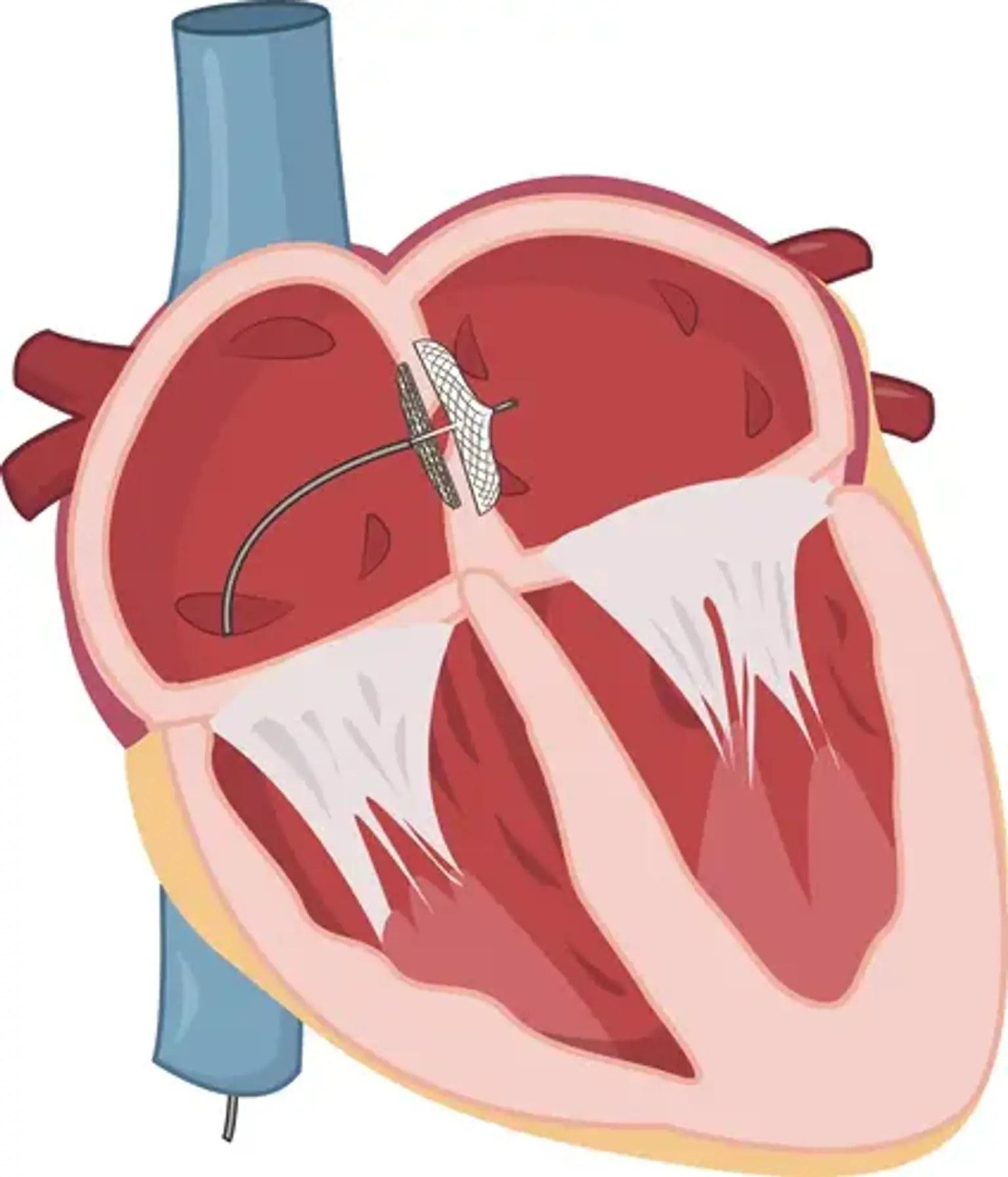
ASD (Atrial Septal Defect) and PFO (Patent Foramen Ovale) are openings in the wall between the upper chambers of the heart. In some patients these openings can place a load on the heart or allow a clot to pass from the right side of the heart to the left side, increasing the risk of stroke.

ASD/PFO closure is the procedure of closing this opening in suitable patients via catheter, without surgery, using a special closure device. The device is usually delivered through a vein in the groin and positioned at the opening in the heart.

The procedure takes approximately 30–60 minutes and most patients can be discharged the following day. In suitable patients it is an effective and safe treatment option without the need for open-heart surgery.

Get in touch with us for an evaluation

Send your appointment request; we'll let you know about the evaluation process suited to your complaint and needs.

Explore Diagnostic Cardiology